Inside the Nose: Exploring the Effects of Microplastics and Nanoplastics on Human Airway Tissue
In This Article
- Mass Eye and Ear researchers show that inhaled microplastics and nanoplastics may contribute to upper airway inflammation, raising new questions about the health effects of chronic environmental exposure
- Using human derived tissue models, the team examined how these ubiquitous particles interact with the nasal lining
- Researchers found that microplastics and nanoplastics interact with the nasal ciliary layer and, in some cases, enter epithelial cells, offering new insight into how these particles may persist in the airway
Microplastics and nanoplastics are now found nearly everywhere in the environment—from oceans and soil to the air people breathe—and growing evidence suggests these tiny plastic particles may pose risks to human health. According to the United States Environmental Protection Agency, microplastics are plastic particles ranging from five millimeters down to one nanometer, while nanoplastics represent the subset smaller than one micrometer. These particles have been detected across diverse ecosystems and have recently been identified in human nasal mucus, with higher levels reported in individuals with conditions such as sinusitis and allergic rhinitis.

Subscribe to the latest updates from Otolaryngology Advances in Motion
Although studies in other mucosal systems, particularly the gut and lungs, have shown that microplastics can trigger inflammatory responses and disrupt epithelial biology, far less is known about their effects on the nasal epithelium. In addition, environmental scientists face significant challenges in quantifying real-world exposure, as many particles are extremely small and difficult to measure accurately. Emerging research also suggests that micro- and nanoplastics may act as carriers for other environmental toxins, known as the “halo effect,” potentially amplifying their biological impact. With the field still in its early stages and major gaps in understanding how these particles behave in human tissue, the nasal mucosa represents a critical but understudied site of investigation.
“The nose is the first place that inhaled particles encounter human tissue,” said Alan D. Workman, MD, MTR, Assistant Professor of Otolaryngology–Head and Neck Surgery at Harvard Medical School and fellowship-trained sinus surgeon at Mass Eye and Ear, whose research focuses on chronic rhinosinusitis and upper-airway inflammatory disease. “Given that exposure is ongoing and these plastics appear to persist within the airway, understanding their potential role as drivers of nasal inflammation represents an important and emerging area of investigation.”
Exploring the human airway in the laboratory
Most current research on microplastics either focuses on detecting plastics in human tissues or relies on animal studies with exposure levels much higher than those humans would typically experience. While these approaches provide important insight, they do not fully capture how microplastics behave in the human airway. Recognizing this gap, Dr. Workman and his team drew on their lab’s long-standing expertise in human nasal epithelial biology to model exposure more accurately using primary human tissue.
To do so, the researchers used patient-derived nasal tissue to create three-dimensional air–liquid interface (ALI) models, in which the apical surface (top) of the cells is exposed to air while the bottom is maintained in culture medium. This setup closely mimics the structure and function of the nasal epithelial lining, enabling physiologically relevant testing of exposures that more closely reflect real-world conditions. Dr. Workman noted that the lab’s extensive experience studying inflammatory airway disease positioned the team well to investigate how micro- and nanoplastics affect epithelial biology.
“Using human tissue and air–liquid interface models lets us interrogate many of the structural and epithelial-derived immune features of the airway in the lab,” Dr. Workman said. “It gives us a more realistic way to study how microplastics and nanoplastics might influence airway inflammation.”
Dr. Workman shares laboratory space with his mentor and now colleague, Benjamin S. Bleier, MD, FACS, Professor of Otolaryngology–Head and Neck Surgery at Harvard Medical School. Their collaboration is supported by shared ideas, methodologies and Institutional Review Board (IRB) protocols, which allow them to utilize human samples—including mucus and surgical tissue—and accelerate specimen collection.
“Our lab is uniquely equipped to study these questions, given our extensive experience with cell culture models,” said Dr. Bleier, who also serves as Director of Endoscopic Skull Base Surgery and Director of Innovation and Commercialization at Mass Eye and Ear. “This provides a rare opportunity to examine microplastic effects directly in human tissue while still allowing findings to be compared with animal models. Insights gained in the nose may also reflect what occurs in the lower respiratory tract, making this approach valuable for broader airway research.”
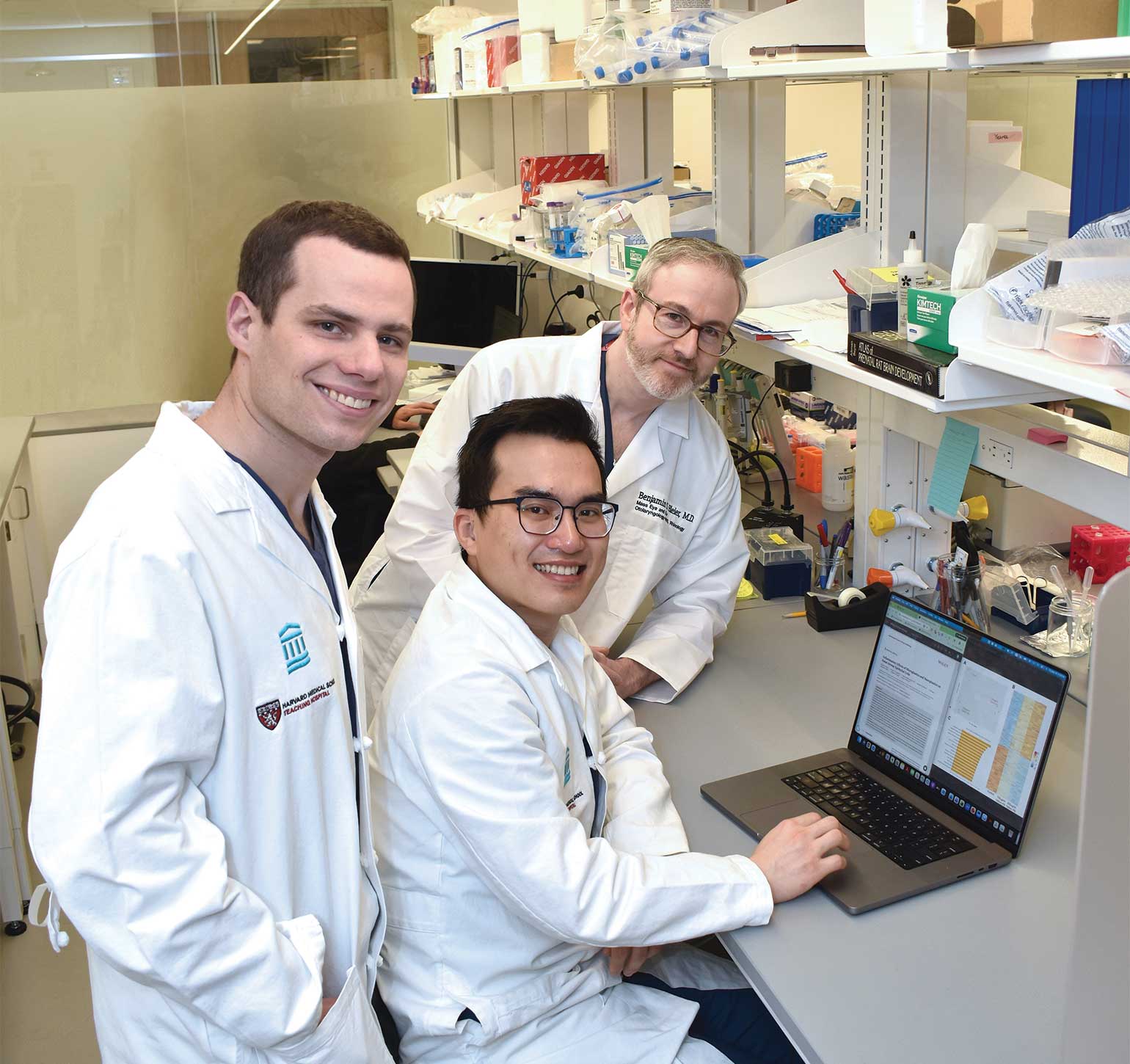
Figure 1
From left to right: Alan D. Workman, MD, MTR; Hoang Bui-Nguyen, MD, PhD (seated); and Benjamin S. Bleier, MD, FACS, collaborating in the laboratory.
Novel findings
The team exposed the apical surface of nasal epithelial cells to polystyrene micro- and nanoplastics over a 10-day period to model repeated daily exposure, with varying particle sizes and concentrations. In addition to studying the cells themselves, researchers analyzed mucus secreted by the ALI cultures to measure soluble inflammatory mediators. Endpoints included inflammatory signaling molecules, gene expression changes, epithelial integrity and cellular ultrastructure. High-resolution confocal microscopy and electron microscopy were used to assess whether particles became trapped within cilia or were taken up into cells.
The findings, published in the International Forum of Allergy & Rhinology, showed that micro- and nanoplastics exposure rapidly activated inflammatory signaling pathways and stress-related genes. Cytokine and gene expression patterns were similar to those associated with allergic rhinitis and chronic rhinosinusitis, with clear effects at the epithelial surface. Exposure also disrupted nasal cilia, although overall cell viability and epithelial barrier function remained intact. Imaging studies indicated that particles could be retained within the ciliary layer and, in some cases, internalized by epithelial cells—an observation the team plans to explore further.
Together, these results suggest that inhaled plastics may contribute to sinonasal inflammation and highlight the utility of nasal epithelial models for studying the health effects of airborne micro- and nanoplastics under chronic, real-world exposure conditions. At the same time, the investigators emphasized that these findings represent early, exploratory data intended to establish foundational mechanistic insights.
Co-first author Hoang C. Bui Nguyen, MD, PhD, a PGY-4 resident in the Harvard Combined Residency Program, led the study’s transcriptomic profiling and computational analyses. He noted that the work is significant because it is the first to demonstrate that micro- and nanoplastics exposure can drive widespread changes in gene regulation within nasal airway epithelial cells. “We found not just isolated inflammatory markers, but broad transcriptomic shifts suggestive of a robust inflammatory response,” Dr. Bui Nguyen explained.
Future directions
According to Dr. Workman, the team is now extending their work to animal models, primary human surgical tissue, and more detailed mechanistic studies to better understand how microplastics and nanoplastics persist in the airway, interact with other environmental toxins, and potentially initiate or exacerbate nasal and lower airway disease. “This is an early and rapidly developing field, where each finding raises new questions about exposure, tissue responses, and long-term health effects,” Dr. Workman shared. “It’s exciting how much opportunity there is to grow this research and begin answering questions that haven’t been explored before.”
A key next step is to define the biological pathways underlying the transcriptomic changes observed in vitro, including whether they are driven by oxidative stress, direct epithelial injury, epigenetic alterations, or other inflammatory signaling mechanisms. The team is also working to better contextualize these findings by comparing experimental exposures with real-world human exposure levels, which remain difficult to quantify due to the extremely small size of many environmental particles.
This forward momentum reflects a collaborative training environment in which shared methods, clinical access, and mentorship are advancing both the science and the next generation of investigators—supporting continued progress from bench models to patient-derived tissue and, ultimately, to clinical translation.
Learn about the Department of Otolaryngology–Head and Neck Surgery