Innovative Zebrafish Model Allows Visualization of Response to Cancer Drugs Over Time at Single-Cell Level and Leads to New Clinical Trial
Key findings
- Researchers at the Massachusetts General Hospital Cancer Center and the Mass General's Department of Pathology have generated immunodeficient zebrafish that are transparent
- The zebrafish allow for direct visualization of tumor growth and response to cancer therapy over time
- Modeling in the zebrafish showed for the first time that olaparib plus temozolomide is a potent therapy against human rhabdomyosarcoma
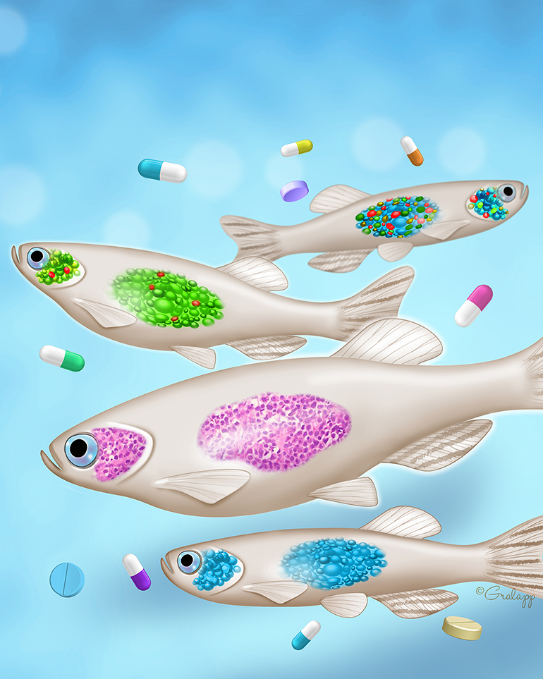
To study the efficacy of cancer drugs prior to human trials, the current standard technique is to graft human cancers into immunodeficient mice. However, this approach has substantial limitations, including the difficulty of imaging individual tumor cells through fur.
Subscribe to the latest updates from Oncology Advances in Motion
In Cell, David M. Langenau, PhD, of the Massachusetts General Hospital Cancer Center and Mass General Department of Pathology, and colleagues report a solution: they have generated immunodeficient zebrafish that are transparent, which allows direct visualization of tumor growth and response to cancer therapy.
Visualizing Engrafted Human Cancer and Therapy Responses in Immunodeficient Zebrafish
Visualizing Single Cells
Cancer researchers often want to visualize tumor cells that have specific functions, such as driving growth, metastasis or therapy resistance. In mice, this is quite difficult; but in the zebrafish, the researchers were able to observe fluorescently labeled human cancer cells under confocal microscopy for more than 28 days. They even quantified tumor growth by counting individual cancer cells.
The zebrafish also enabled visualization of cancer cell behavior over extended periods of time. As a test case the researchers imaged human rhabdomyosarcoma cells and observed for the first time in vivo that there are three functionally distinct types.
Testing a Combination Therapy
The functional diversity of the rhabdomyosarcoma cells provided a rationale for the first preclinical trial of a dual-mechanism treatment for rhabdomyosarcoma. The researchers engrafted the zebrafish with human alveolar rhabdomyosarcoma, embryonal rhabdomyosarcoma or, as a control, Ewing's sarcoma. They then dosed the fish with olaparib (a PARP inhibitor), temozolomide (a DNA-damaging agent) or the combination.
Over 28 days, the monotherapies showed little effect on tumor growth, but combination treatment led to potent tumor regression in all three tumor types. These results were recapitulated in patient-derived xenografts grown in mice, providing additional preclinical rationale for opening a phase I clinical trial that is now accruing patients at the Mass General Cancer Center and Dana Farber Cancer Institute.
Toward the Future
The researchers envision using the zebrafish to:
- Visualize additional hallmarks of cancer, such as cancer-associated vascular networks
- Conduct large-scale, high-throughput drug screening
- Grow a patient's own tumor to identify the most appropriate clinical trial for that patient
view original journal article Subscription may be required
Learn more about the Langenau Lab
Refer a patient to the Mass General Cancer Center