Ultrasound with Micropuncture Needle Reduces Vascular Access Complications in Cardiac Electrophysiological Procedures
In This Article
- In this poster presented at Clinical Research Day at Mass General, this study seeks to reduce vascular access complications after electrophysiological procedures, which are among the most common
- Previous studies in the field evaluated real-time ultrasound (US) guidance and micropuncture needle for vascular access separately but not in combination
- The researchers sought to determine whether the use of US to guide vascular access along with a 21 G micropuncture needle would reduce major vascular complication
- They found that direct US visualization in combination with micropuncture needle significantly reduces major vascular complications, compared to a standard 18 G needle without ultrasound guidance
Background
Vascular access complications are among the most common complication encountered after electrophysiological procedures. Previous studies in the field evaluated real-time ultrasound (US) guidance and micropuncture needle for vascular access separately but not in combination.
Subscribe to the latest updates from Cardiovascular Advances in Motion
Purpose
In this study, the researchers led by Chee Yuan Ng, MD, FACC, cardiologist at Massachusetts General Hospital, sought to determine whether the use of US to guide vascular access along with a 21 G micropuncture needle would reduce major vascular complication.
Methods
- 182 consecutive patients undergoing cardiac electrophysiological procedures were included in the study.
- Vascular access was performed under direct US visualization of the 21 G needle. When prescribed, anticoagulation was continued during the procedure.
- The control group consists of 162 consecutive patients who underwent vascular access with a standard 18 G needle.
- Major vascular complications were defined as haematoma requiring intervention, arterial pseudoaneurysm and arterio-venous fistula.
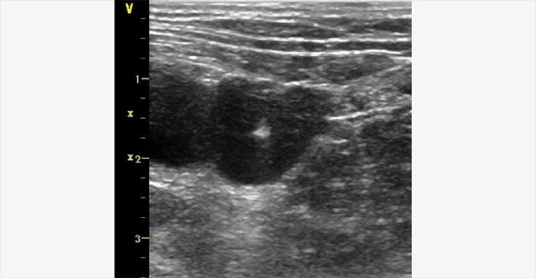
Fig. 1: Direct Visualization of the 21 G Micropuncture Needle Tip Entering the Femoral Vein with Ultrasound Imaging
Results
- The most common procedure performed was ablation for atrial fibrillation, which accounts for 54% of all procedures.
- 12% of procedures were ablation for ventricular tachycardia and other procedures accounted for the remaining 34% of the procedures.
- 0/182 or 0% major vascular complications were identified in the study group, compared to 6/162 or 3.7% in the control group (p=0.01).
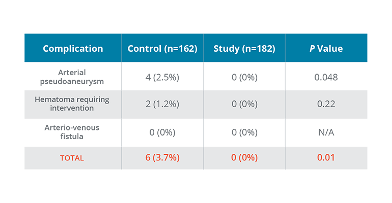
Fig. 2: Table Comparing Study Complications to Control Complications
0% major vascular complications were identified in the study group compared to 3.7% in the control group.
Conclusion
- Direct US visualization, in combination with micropuncture needle, significantly reduces major vascular complications, compared to a standard 18 G needle without ultrasound guidance.
- The results were presented at the European Heart Rhythm Association Congress 2017.
Authors
Chee Yuan Ng, MD, Guy Rozen MD, Yitschak Biton MD, Jordan Leyton-Mange MD, Conor Barrett MBBCh, BAO.
Massachusetts General Hospital, Harvard Medical School, Boston, MA.
Learn more about the Telemachus & Irene Demoulas Family Foundation Center for Cardiac Arrhythmias
Learn more about Dr. Barrett’s research