Developing a Personalized Approach for Targeted Radiotherapy of Liver Cancers
In This Article
- Investigators at Massachusetts General Hospital published findings demonstrating a computational method that enhances clinical decision-making for delivering radiation in liver cancer
- The model simulates the movement of radioactive microspheres through liver vasculature to optimize the targeted release of radiation in the tumor
- The microsphere dosimetry (MIDOS) model enables a personalized approach to transarterial radioembolization (TARE) planning to maximize radiotherapy effectiveness and avoid adverse outcomes
Massachusetts General Hospital researchers are developing methods to enhance the safety and effectiveness of radiation therapy for liver cancers. Their microsphere dosimetry (MIDOS) model allows clinicians to fine-tune conditions critical for the targeted delivery of radiation to tumor tissue.
Subscribe to the latest updates from Oncology Advances in Motion
"Although traditional methods used to perform external radiotherapy are highly precise, optimizing factors under in vivo conditions makes this process significantly more complicated," says Alejandro Bertolet, PhD, head of B-Lab (Dosimetry and Modeling for RPT) at Radiation Oncology at Mass General and assistant professor of Radiation Oncology at Harvard Medical School. "This research represents a first step in applying computational techniques to increase the precision of personalized targeted radiotherapy."

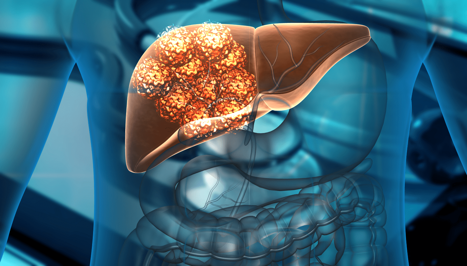
Figure 1
3D illustration of human liver cancer cell growth.
Enhancing Techniques for the Targeted Treatment of Liver Cancers
Liver cancers are the third-leading cause of cancer-related death worldwide. In early-stage tumors, resection of the tumor and surrounding tissue is the treatment of choice. For patients with later-stage, unresectable tumors who are not transplant candidates, treatment focuses on palliative measures capable of downstaging the tumor to enable resection.
Treatment options for unresectable tumors include transarterial therapies, which involve the delivery of small spherical particles (microspheres) through a catheter inserted via the hepatic artery. The microspheres lodge in tumor blood vessels, where they obstruct (embolize) the flow of oxygenated blood, resulting in cancer cell death and tumor shrinkage. This technique has been successfully expanded to allow the delivery of a radioactive compound via transarterial radioembolization (TARE).
Given its therapeutic value, TARE applications have evolved rapidly in recent years. However, Dr. Bertolet suggests that this evolution has outpaced methods to control for uncertainties associated with this targeted treatment. "The rationale for this therapy is delivery of the highest radiation dose to a tumor without affecting normal tissue or causing other significant off-target effects," he advises.
"Our experience with the high degree of accuracy in traditional radiation oncology procedures revealed aspects of TARE planning that could be enhanced," explains Bertolet. "This led us to create a model that can augment the planning process in order to maximize the probability of therapeutic success."
Creating an Atlas of Liver and Tumor Blood Vessels
TARE planning begins with the injection of contrast dye into the liver and tumor to allow visualization of blood vessels via diagnostic computed tomography angiography. This establishes a baseline vascular map of both areas that offers possible catheter-access points for TARE. Afterward, the access points are used to administer a biodegradable radiopharmaceutical agent that mimics the characteristics of the radioactive microspheres.
Introducing these agents allows accurate tracking of their distribution throughout the liver and tumor(s), as well as possible transitions to the heart, lungs, and abdomen. This simulation of microsphere movement offers critical information about possible pathways the eventual microspheres used for treatment could follow once administered, as well as data that allow calculations of two important parameters:
- Lung shunt fraction (LSF) estimates the absorbed dose of a radioactive isotope in the lung relative to the total between the lung and liver.
- Tumor-to-normal ratio (TNR) estimates the difference in radiation taken up by the tumor relative to non-tumor hepatic tissues.
In terms of the LSF, tumors can induce abnormalities in liver vasculature that result in shunts capable of redirecting arterial blood flow through the hepatic vein to the heart and into the lungs. For TARE planning, the LSF and TNR offer insights into whether alternative catheter-entry points should be considered.
Dr. Bertolet notes that while this information provides an essential patient-specific roadmap, it fails to account for other TARE-related uncertainties appropriately, including:
- Divergence in the physical properties between the radioactive microspheres and radiotherapeutic agents used for the simulations
- Inconsistencies in the quality and definition of angiographic imaging
- Differences in microsphere material and its impact on their radiation uptake and release, as well as how they cluster and lodge within blood vessels
- The rate of decay in the specific radioisotope being delivered
"Although we cannot control aspects of the physiological environment, we can account for many factors by providing information about other essential components of the system that directly affect TARE outcomes," he says.
Building Models Capable of Optimizing Clinical Decision-Making
Real-time radiation delivery introduces numerous variables into an equation designed to protect the patient and maximize the targeted effect of the radiation. The MIDOS model employs a computational representation of liver vasculature generated from data from reference adult males and adult females by collaborators at the University of Florida. Tumors with an algorithm-generated vascular network can then be placed at any location within the construct.
MIDOS aims to predict the amount of radiation that will be delivered to tumors, normal liver tissue, and the lungs according to the following user-provided parameters:
- Amount of radiation to be delivered
- Microsphere characteristics (size and material)
- Catheter-entry point
- Delivery timing (to account for radioactive decay)
- Estimated LSF
The team's simulations showed that radiation is more likely to be delivered to tumors with higher degrees of vascularity versus normal tissue (a higher TNR). Similarly, they found that microsphere type and radiation load significantly affected both the TNR and LSF, suggesting an ability to optimize treatment by selecting the right "delivery vehicle."
"MIDOS successfully replicates clinically relevant TARE scenarios, allowing us to evaluate decisions based on different treatment options," says Dr. Bertolet.
The next step is to capitalize on advanced imaging technologies to generate patient-specific representations of the liver and tumor and refine the methods used to simulate microsphere movement.
"The model's adaptability and flexibility support levels of radiotherapy optimization that will make personalized approaches to treating liver cancers a reality."
An Environment That Fosters Discovery
Dr. Bertolet emphasizes the Mass General ecosystem as invaluable to making MIDOS a reality and a primary motivation in establishing a lab in the Department of Radiation Oncology.
"The encouragement I've received in the form of commitments and collaborations for this work has been incredible," he says. "The success of this research is a direct result of the amazing support from other investigators and clinicians, especially Eric Wehrenberg-Klee, MD, from Interventional Radiology, a world-class expert on these treatments, besides the entire Division of Nuclear Medicine and Molecular Imaging."
Proximity to the Boston area also has its benefits, including having Boston Scientific as neighbors. As manufacturers of the microspheres used for TARE, their willingness to collaborate was enormously beneficial.
"We absolutely could not have accomplished this without their insight and experience," Dr. Bertolet says.
He adds that the value Mass General places on research makes it unique, especially in his field.
"As a medical physicist, my ability to be 100% dedicated to research in a hospital setting is very rare. This is a once-in-a-lifetime opportunity to exclusively focus on translating science into therapies that can have an immediate impact on patients."
Learn more about our Liver Cancer Treatment Program
Discover more about the Mass General Cancer Center